What is an Ultrasound?
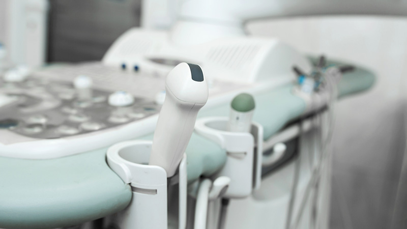
An ultrasound is safe and painless, and produces pictures of the inside of the body using sound waves. Ultrasound imaging, also called ultrasound scanning or sonography, involves the use of a small transducer (probe) and ultrasound gel placed directly on the skin. High-frequency sound waves are transmitted from the probe through the gel into the body. The transducer collects the sounds that bounce back and a computer then uses those sound waves to create an image. Ultrasound examinations do not use ionizing radiation (as used in x-rays), thus there is no radiation exposure to the patient. Because ultrasound images are captured in real-time, they can show the structure and movement of the body’s internal organs, as well as blood flowing through blood vessels.
Ultrasound imaging is a noninvasive medical test that helps physicians diagnose and treat medical conditions.
Conventional ultrasound displays the images in thin, flat sections of the body. Advancements in ultrasound technology include three-dimensional (3-D) ultrasound that formats the sound wave data into 3-D images.
A Doppler ultrasound study may be part of an ultrasound examination.
Doppler Ultrasound is a special ultrasound technique that allows the physician to see and evaluate blood flow through arteries and veins in the abdomen, arms, legs, neck and/or brain (in infants and children) or within various body organs such as the liver or kidneys.
An Ultrasound can help provide physicians with information on:
Ultrasound examinations can help to diagnose a variety of conditions and to assess organ damage following illness.
Ultrasound is used to help physicians evaluate symptoms such as:
- Pain
- Swelling
- Infection
Ultrasound is a useful way of examining many of the body’s internal organs, including but not limited to the:
- heart and blood vessels, including the abdominal aorta and its major branches
- liver
- gallbladder
- spleen
- pancreas
- kidneys
- bladder
- uterus, ovaries, and unborn child (fetus) in pregnant patients
- eyes
- thyroid and parathyroid glands
- scrotum (testicles)
Doppler ultrasound images can help the physician to see and evaluate:
- blockages to blood flow (such as clots)
- narrowing of vessels
- tumors and congenital vascular malformations
- less than normal or absent blood flow to various organs
- greater than normal blood flow to different areas which is sometimes seen in infections
What to expect during your Ultrasound
- For most ultrasound exams, you will be positioned lying face-up on an examination table that can be tilted or moved. Patients may be turned to either side to improve the quality of the images.
- After you are positioned on the examination table, the sonographer will apply a warm water-based gel to the area of the body being studied. The gel will help the transducer make secure contact with the body and eliminate air pockets between the transducer and the skin that can block the sound waves from passing into your body. The transducer is placed on the body and moved back and forth over the area of interest until the desired images are captured.
- There is usually no discomfort from pressure as the transducer is pressed against the area being examined. However, if scanning is performed over an area of tenderness, you may feel pressure or minor pain from the transducer.
- Doppler sonography is performed using the same transducer.
Preparing for your Ultrasound
- Please arrive 15 minutes before your appointment time.
- You should wear comfortable, loose-fitting clothing for your ultrasound exam. You may need to remove all clothing and jewelry in the area to be examined.
- You may be asked to wear a gown during the procedure.
- Preparation for the procedure will depend on the type of examination you will have. For some scans your doctor may instruct you not to eat or drink for as many as 12 hours before your appointment. For others you may be asked to drink up to six glasses of water two hours prior to your exam and avoid urinating so that your bladder is full when the scan begins.
Results
Your Ultrasound results will be reported to your referring physician within 1-2 business days.
Your referring physician will contact you with your results.