Delivers Superior Diagnostic Accuracy
Provision Diagnostic Imaging became the first imaging center in East Tennessee to offer Cardiac PET/CT with N-13 Ammonia, which provides the most efficient, precise, and advanced imaging for diagnostic and therapeutic purposes. As part of our mission to embrace innovative healthcare solutions, we recognize the Cardiac PET/CT scan as one of the most distinguishing diagnostic tools in the cardiovascular industry to assess myocardial blood flow, as well as microvascular and triple vessel disease.
Joint Position Statement
American Society of Nuclear Cardiology
Society of Nuclear Medicine and Molecular Imaging
Regarding Clinical Indications for Rest-Stress Myocardial Perfusion Imaging (MPI):
“Cardiac PET is a first line preferred test for patients with known or suspected coronary artery disease (CAD) who meet appropriate criteria for a stress imaging test and are unable to complete a diagnostic level exercise stress imaging study. There are no clinical scenarios where PET should not be considered a preference test for patients who meet appropriate criteria for a stress imaging test and who require pharmacological stress.”
ASNC/SNMMI POSITION STATEMENT
What is Cardiac PET/CT with N-13 Ammonia?
PET is positron emission tomography, which is an imaging scan technique that uses radioactive substances known as radiotracers to visualize and diagnose a variety of diseases. In the Cardiac PET scan, the focus is specific to Cardiac, which refers to heart disease. Ammonia N-13 is a vital radiotracer utilized specifically for Cardiac PET imaging. The CT scan stands for computerized tomography, which combines a series of x-ray images taken from different angles around the body and uses computer processing to create cross-sectional images (slices) of the bones, blood vessels and soft tissues inside the body. The combination PET technique and CT imaging data provide comprehensive scans of anatomy and function at a relatively low radiation dose, allowing the most precise visibility to the heart muscle function and blood flow, referred to as myocardial perfusion image (MPI).
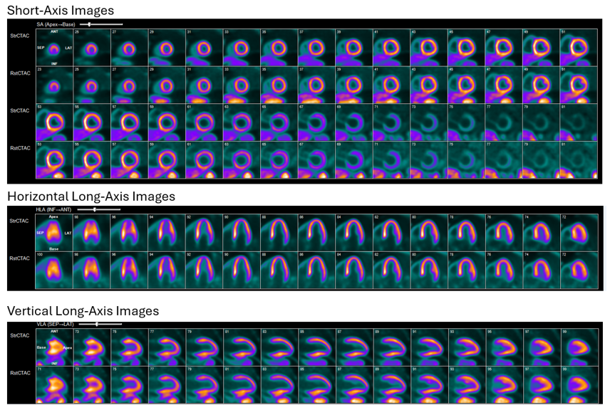
N-13 Ammonia Cardiac PET/CT Images Comparing Myocardial Perfusion on both Rest and Stress Phases
How Does Cardiac PET/CT Work?
When combining PET and CT technologies with N-13 Ammonia, a 3-D image shows the MPI, myocardial perfusion image, which illustrates the function of the heart muscle and absolute quantification of myocardial blood flow in tissue, during stress and rest conditions. This plays a significant role in diagnostic and therapeutic decision making in the field of cardiac disease.
Electrical conductors called electrodes are placed strategically as stickers on your chest to monitor the electrical activity of your heart during both a resting and stressed state. The stress state of the heart is achieved when the heart is pumping hard and can be produced through exercise on a treadmill or with medication that makes the heart respond as if you were physically exercising. A small radiopharmaceutical agent injected into your IV line allows the PET camera to capture pictures of your heart during these two phases: the resting phase and stressed phase. An accurate assessment of your blood flow is then made when comparing the two phases.
While there are multiple modalities to detect coronary heart disease (CAD), N-13 Ammonia yields the highest quality image in the least amount of time compared to SPECT (Single-Photon Emission Computed Technology) and Cardiac PET/CT using Rubidium. SPECT and PET use different radiotracers, where SPECT radiotracers measure gamma rays; the decay of the radiotracers used with PET produce small particles called positrons. A SPECT scan may not detect vascular disease at the micro-circulatory level of the coronary circulation, or early stages of coronary artery disease when there are no symptoms, yet a PET scan can detect early functional abnormalities of the coronary circulation, which may be a precursor of ensuing coronary artery disease.
What are the Benefits of Cardiac PET/CT with N-13 Ammonia?
- Improved Diagnostic Accuracy:
- N-13 Ammonia has high sensitivity and accuracy for detecting coronary heart disease.
- Smaller Correction Factors:
- N-13 has more reliable quantification, resulting in fewer correction factors with myocardial blood flow.
- Better Extraction Fraction and Retention:
- N-13 extraction is close to 100% at rest and stress (0.95-0.99); and has the highest retention of FDA approved tracers (0.50-0.90)
- Lower radiation exposure compared to SPECT MPI
- Shorter image time for patient compared to conventional SPECT MPI (35 min vs. 3-4 hours)
- Quantitation of myocardial blood flow, which is valuable in diagnosing triple vessel and microvascular disease.
- Improved risk stratification and patient management
Who is a Candidate for Cardiac PET/CT?
- For patients who have symptoms and/or risk factors for heart disease.
- For patients with prior suboptimal or equivocal imaging or stress exams
- For patients who are morbidly obese, in whom both SPECT MPI and Stress Echocardiography routinely can be inconclusive.
- For patients with BMI > 30-35 kg/M2 because body habitus that may affect image quality (obesity, large breasts, or implants, etc.)
- For heart failure patients who need new workup because they are not qualified for heart Cath
- For young patients with known CAD, who will require repeat testing over their lifetime.
- For patients where myocardial blood flow quantitation is required.
- For patients concerned with multi-vessel disease with low ejection fraction
- For high-risk patients: diabetes, chronic kidney disease, multi-vessel or left main disease.
What to Expect on your Cardiac PET/CT appointment
- Please arrive 30 minutes prior to your appointment.
- Wear warm and comfortable clothing you may keep on during your test.
- Bring a list of your medications and be aware of special instructions if you are diabetic.
- Do not eat or drink anything except water for 4-6 hours prior to your test.
- Do not have caffeine, chocolate, or decaffeinated products at least 12 hours prior to your test.
- Check with your physician before discontinuing any medications.
- Your PET CT exam should last approximately 1 hour. If longer, this should not be of concern.
- Let your healthcare professional know if you experience any chest pain, shortness of breath, palpitations, headache, dizziness, lightheadedness, or a sloshing feeling during the test.
- Results and images will be sent to your referring physician provider.